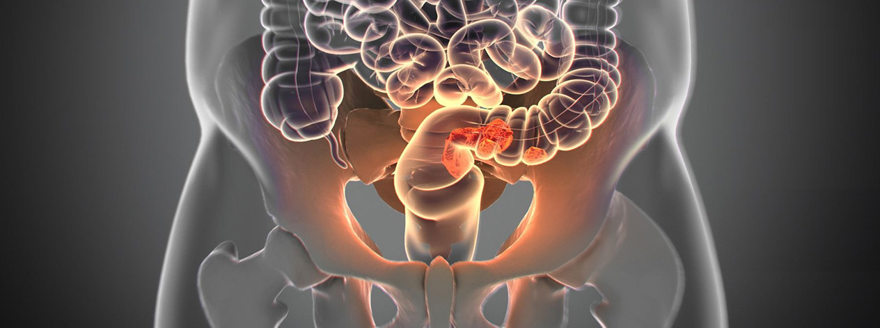
Rectal cancer
The colon and rectum are parts of the body’s digestive system, and together with the anal canal and anus form a long, muscular tube called the large intestine. The colon is the first 6 feet of the large intestine. The rectum and anal canal form the last several inches. The large intestine ends at the anus, which is the opening to the outside of the body.
Although cancers of the colon and rectum are often referred to jointly as “colorectal cancer,” treatment approaches differ and are discussed separately. Anal cancer is a distinct type of cancer (the term “colorectal cancer” does not include anal cancer), and is also discussed separately.
Adenocarcinoma is the most common type of cancer that originates in the cells that line the rectum or large intestine. It accounts for over 90-95% of cancers originating in the rectum. Other types of cancer including carcinoid and leiomyosarcoma also originate in the rectum, but are not referred to as rectal cancer. This treatment overview deals only with adenocarcinoma of the rectum, which will be referred to as rectal cancer.
The treatment of rectal cancer may involve several physicians, including a gastroenterologist, a surgeon, a medical oncologist, a radiation oncologist, and/or other specialists. Care must be carefully coordinated between the various treating physicians involved in management of your cancer.
STAGING
In order to understand the best treatment options available for treatment of rectal cancer, it is important to first determine where the cancer has spread in the body. The initial spread of rectal cancer occurs circumferentially around the rectum and laterally into the adjacent fat and muscles. Rectal cancer can then invade nearby organs and spread through the lymph and blood systems. Rectal cancer cells may spread via the blood throughout the body to the liver, lungs and other organs.
Determining the stage of the cancer or the extent of the spread requires a number of tests and is ultimately confirmed by surgical removal of the cancer and exploration of the abdominal cavity.
Computerized Tomography (CT) Scan: A CT scan is a technique for imaging body tissues and organs, during which X-ray transmissions are converted to detailed images, using a computer to synthesize X-ray data. A CT scan is conducted with a large machine positioned outside the body that can rotate to capture detailed images of the organs and tissues inside the body.
Magnetic Resonance Imaging (MRI): MRI uses a magnetic field rather than X-rays, and can often distinguish more accurately between healthy and diseased tissue. MRI gives better pictures of tumors located near bone than CT, does not use radiation as CT does, and provides pictures from various angles that enable doctors to construct a three-dimensional image of the tumor.
Colonoscopy: A colonoscopy may be used to identify whether a second cancer is present in the colon or rectum prior to surgery. During a colonoscopy, a long flexible tube that is attached to a camera is inserted through the anus, allowing physicians to examine the internal lining of the colon and rectum for polyps or other abnormalities. The physician may perform a biopsy during a colonoscopy in order to collect samples of suspicious tissues or cells for closer examination.
Endorectal Ultrasound (EUS): Endorectal ultrasound (EUS) involves the use of a special probe that is inserted into the rectum to help determine the thickness of the cancer. By determining the thickness of the cancer, EUS can help determine the stage.
SURGERY
Upon completion of the clinical staging evaluation, surgery is performed to remove the cancer, along with part of the normal adjacent tissues of the rectum. Surgery also helps to further determine the level of spread within the rectal wall and abdomen. The type of surgery performed depends on the size and the location of the cancer. For more information, go to Surgical Management of Rectal Cancer.
Some patients may be treated with radiation therapy and/or chemotherapy prior to surgery. Giving these treatments before surgery may reduce the risk of cancer recurrence and help to shrink the cancer prior to surgery.
Following surgical removal of rectal cancer, a final “pathologic” stage will be given. This is based on extent of spread of cancer after looking at the removed tissue under a microscope. All new treatment information concerning rectal cancer is categorized and discussed by the stage. In order to learn more about the most recent information available concerning the treatment of rectal cancer, click on the appropriate stage.
Stage I: Cancer is confined to the rectum.
Stage II: Cancer may penetrate the wall of the rectum into the surrounding fat or muscles or other adjacent organs, but does not invade any local lymph nodes.
Stage III: Cancer invades one or more of the local lymph nodes, but has not spread to other distant organs.
Stage IV: Cancer has spread to distant locations in the body, which may include the liver, lungs, bones or other sites.
Recurrent/Relapsed: The rectal cancer has progressed or returned (recurred/relapsed) following an initial treatment
Copyright © 2020 Omni Health Media Rectal Cancer Information Center. All Rights Reserved.